newsletter
Get useful insight and advice in your inbox.
Thank you! Your submission has been received!
Oops! Something went wrong while submitting the form.
-8.png)
Fuchs' dystrophy and cataracts often arrive together, because both worsen with age, and operating on one without a plan for the other can cost you the cornea. Cataract surgery stresses the corneal endothelium, the delicate one-cell-thick layer that keeps your cornea clear. If that layer is already compromised by Fuchs', routine surgery can tip it into failure. So the surgeon's plan, technique, and even the viscoelastic gel used inside the eye all change. Fuchs' endothelial dystrophy affects roughly 4 percent of US adults over 40, and most carriers do not know it until a cataract consult turns up guttata on the slit lamp (AAO EyeWiki). This guide explains the condition, the three surgical paths, and exactly how an experienced surgeon spares the endothelium.
The cornea is the clear front window of the eye. It stays clear because a single layer of cells on its inner surface, the corneal endothelium, continuously pumps fluid out of the cornea. These cells do not regenerate. You are born with a finite supply, and the count declines slowly over a lifetime. When the count falls too low, the pump fails, fluid builds up, and the cornea swells and clouds.
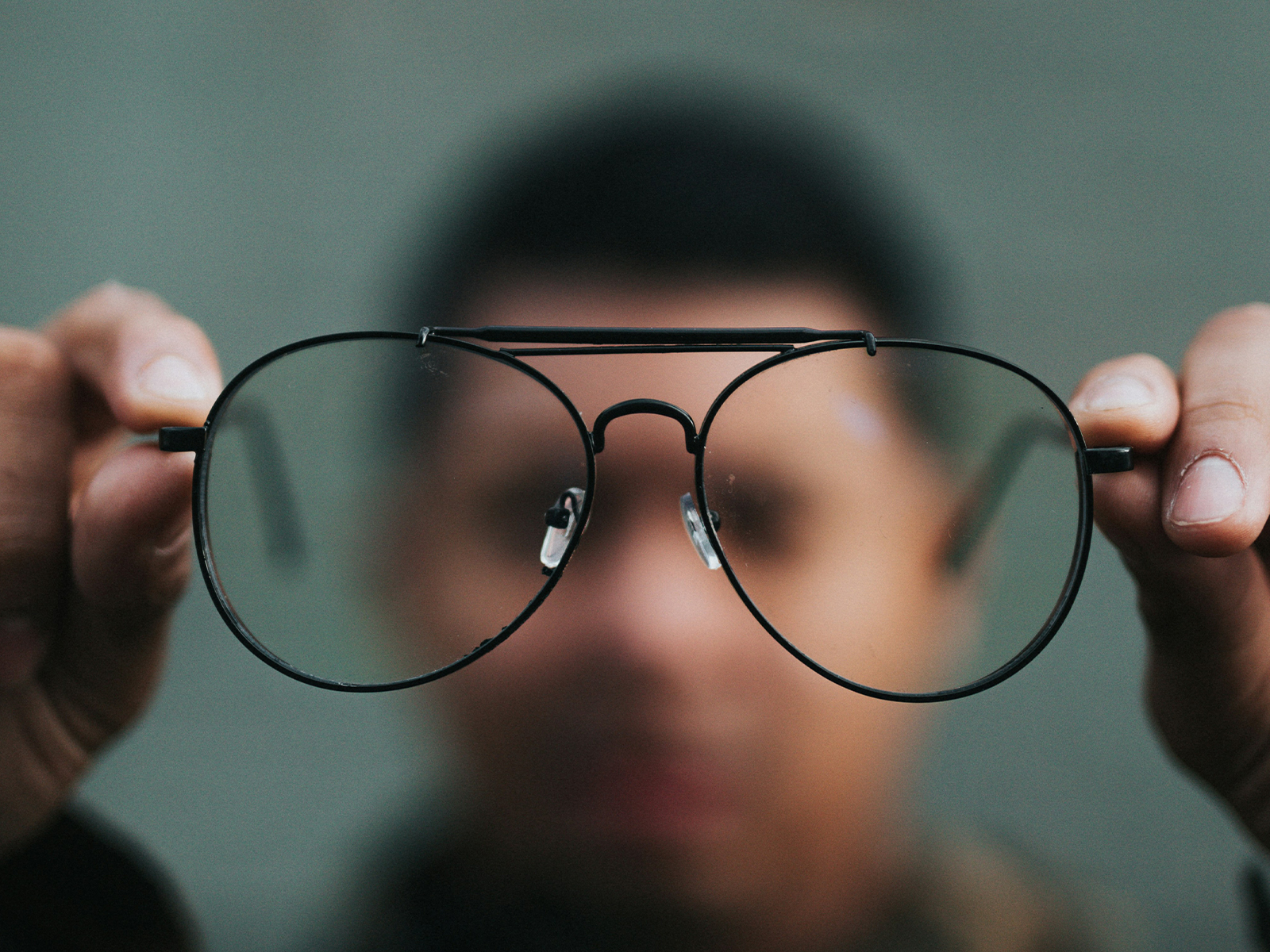
Fuchs' dystrophy is a genetic condition in which the endothelial cells produce abnormal bumps of excess basement membrane called guttata (also described in the literature as Hassall-Henle bodies when peripheral). At the slit lamp, a surgeon sees these as a beaten-metal appearance on the back of the cornea. Spotting guttata at a cataract evaluation is frequently the moment a patient first learns they have Fuchs'.
We measure the health of the endothelium with specular microscopy, which photographs and counts the cells. A healthy adult cornea has roughly 2,000 to 3,000 cells per square millimeter. As the count drops toward 1,000 and below, the cornea's reserve to withstand the stress of surgery shrinks. The cell count, along with how much the cornea is already swollen, drives the whole surgical plan.
Fuchs' progresses in stages. Early on, guttata are present but the patient has no symptoms. Next comes morning blur: the cornea swells overnight when the eyelids are closed and clears as the day's open-eye evaporation pulls fluid out. As the disease advances, the blur lasts longer into the day, then becomes constant, and eventually painful epithelial blisters can form. Recognizing where you sit on that curve is central to timing surgery.
Phacoemulsification, the ultrasound energy used to break up and remove the cataract, releases energy and fluid turbulence inside the eye that costs every eye some endothelial cells. A healthy cornea absorbs that loss easily. A Fuchs' cornea has less reserve to spare.
If the endothelial count is already low, the cell loss from surgery can push it below the threshold where the pump keeps up. The cornea then stays swollen and cloudy after surgery, a state called corneal decompensation, which may require a corneal transplant to fix. We state this plainly: cataract surgery in a Fuchs' eye can hasten corneal failure. The job of the plan is to make that outcome as unlikely as possible. Read more about dry eye and cataract surgery results, which often coexists and compounds surface symptoms.
Before surgery we assess central corneal thickness with pachymetry (a thicker cornea signals more swelling), the endothelial cell count and morphology on specular microscopy, and the practical question of how long your vision stays blurry in the morning. Together these tell us whether the cornea can safely tolerate cataract surgery alone or whether it needs help.
There is no single right answer for every Fuchs' eye. The decision is individualized, and it usually lands on one of three paths.
When guttata are mild, the cell count is healthy, the cornea is not swollen, and morning blur is minimal or absent, cataract surgery alone, performed with endothelial protection technique, is appropriate. Many patients with early Fuchs' do exactly this and never need anything more.
For an intermediate eye, we may do the cataract surgery first with maximal endothelial protection, then watch how the cornea responds. If it stays clear, no further surgery is needed. If it decompensates over the following months, we add a corneal transplant (DMEK) as a second, separate procedure. This staged approach avoids transplanting a cornea that might have done fine on its own.
When the Fuchs' is moderate to severe, the cornea is already swelling, or the cell count is low enough that decompensation is likely, we combine the cataract surgery and the DMEK transplant in a single operation. This is called the triple procedure: cataract removal, IOL implantation, and DMEK together. It saves the patient a second surgery and a second recovery when transplant is clearly going to be needed.
DMEK is a partial corneal transplant. Instead of replacing the whole cornea, the surgeon removes only the failed inner layer (the endothelium and its underlying Descemet's membrane) and replaces it with an ultra-thin donor graft of healthy endothelial cells and Descemet's membrane. The graft is unfolded inside the eye and held against the back of the cornea with an air or gas bubble until it adheres.
An older technique, DSAEK, transplants a thicker layer that includes some donor stroma. DMEK transplants only the Descemet's membrane and endothelium, which is thinner and more anatomically natural. For Fuchs', DMEK has largely become the preferred approach because it generally delivers sharper vision, faster visual recovery, and a lower rejection rate than DSAEK (Cornea Society). It is more technically demanding to perform, which is one reason surgeon experience matters.
DMEK recovery is defined by two things: positioning and patience. In the first day or two you keep your face up so the bubble presses the graft into place. Vision then clears gradually over weeks to a few months as the new cells pump the cornea dry. This is slower than a standalone cataract recovery, and we set that expectation clearly up front.
Whether or not a transplant is planned, the technique used during the cataract portion determines how many precious endothelial cells survive. This is where surgeon experience earns its keep, and the patient deserves to know the named tools.
Viscoelastic is the protective gel injected into the eye during surgery. A dispersive viscoelastic such as Viscoat or Healon EndoCoat coats and clings to the endothelium, forming a shield that absorbs ultrasound energy and shock waves before they reach the cells. In a Fuchs' eye, we re-coat the endothelium repeatedly through the case to maintain that shield.
The soft-shell technique layers a dispersive viscoelastic against the cornea and a cohesive viscoelastic beneath it, combining the protective coating of one with the space-maintaining stability of the other. It is a standard endothelial-sparing maneuver in compromised corneas.
We lower the fluid flow and bottle height so there is less turbulence inside the eye, and we use torsional (oscillating) ultrasound and chopping techniques that remove the cataract with less total energy. Less energy and less turbulence mean fewer cells lost.
Endothelial cell loss correlates with how efficiently the cataract is removed, which correlates with experience. A high-volume surgeon familiar with these eyes works with less energy and less manipulation. We do not claim to be the best, but we do operate on Fuchs'-complex eyes routinely, and that familiarity is protective.
For eyes that need DMEK, the cornea specialist relationship is part of standard high-quality care, not a hand-off. Whether the transplant is performed in-house or with a corneal colleague, the cataract and corneal plans are made together so the IOL choice, the timing, and the technique all align. We handle and co-manage these cases as a normal part of our practice. You can read our broader guidance on cataract surgery with concurrent eye conditions.
Multifocal and trifocal lenses split light to create multiple focal points, which depends on a perfectly clear cornea to deliver good image quality. In moderate to severe Fuchs', the cornea may never be flawlessly clear, and any residual haze degrades the multifocal image and worsens glare. For that reason these lenses are usually not recommended in moderate to severe Fuchs', though the decision is nuanced rather than an absolute rule. We make the call based on your specific corneal status.
A monofocal lens is the dependable default and asks the least of the cornea. An extended depth of focus lens such as the Vivity EDOF can be reasonable in milder Fuchs' with a healthy endothelium, because it is more forgiving of imperfect optics than a trifocal while still extending range.
Toric lenses correct astigmatism, but in a triple-procedure eye the DMEK can slightly change the cornea's shape and therefore the astigmatism as it heals. We are cautious committing to a fixed toric correction before the cornea has settled, because the target can move.
This uncertainty is exactly why the Light Adjustable Lens (LAL) is a strong option for Fuchs' eyes, especially triple-procedure eyes. The LAL is implanted at a best estimate, and its power, including its astigmatic correction, is fine-tuned with ultraviolet light treatments after the cornea has fully recovered and the final refraction is known. When the cornea's shape is a moving target, a lens you can adjust after the dust settles is the right tool. A broader primer is in our intraocular lens options guide.
After cataract surgery alone, you go home with a shield and start drops; vision is often functional the next day. After a triple procedure, you also begin strict face-up positioning to keep the DMEK bubble in place, and your vision will be hazy while the cornea is still swollen.
Triple-procedure patients follow a positioning routine in the first days and a steroid drop schedule that tapers over months to prevent graft rejection. Cataract-alone patients follow the standard, shorter drop course. Our general cataract recovery timeline applies to the cataract portion.
This is the single most important expectation to set. The cataract portion clears in days. The DMEK portion clears over weeks to months as the donor cells dehydrate the cornea. Patience in the first month is normal and expected, not a sign that something is wrong.
A DMEK graft loses endothelial cells gradually over the years, as all corneas do, and a minority of patients eventually need a repeat graft years later. This is a known, manageable part of the long arc, and we discuss it honestly rather than implying a transplant is permanent and final.
Medicare and most commercial plans cover medically necessary cataract surgery and medically necessary DMEK, including when combined as a triple procedure for documented Fuchs' decompensation. These are covered medical services when the clinical indication is documented.
As with any cataract surgery, Medicare covers the operation and a standard monofocal lens, but the refractive upgrade portion of a premium lens such as a Light Adjustable Lens or a toric remains a patient-pay cost. We never represent a premium lens upgrade as covered.
The figures and coverage rules described here are 2026 estimates and vary by plan and by whether your deductible is met. Confirm your specific responsibility with your plan before scheduling.
A subspecialty practice experienced with Fuchs'-complex eyes will answer each of these in terms specific to your cornea. You can read more about our practice and our approach to complex cases, review our overview of complex cataract surgery and femtosecond-assisted cataract surgery, and read about potential complications of cataract surgery and what to expect after cataract surgery. This article was prepared by the surgical team at Modern Cataract Surgery, including Brent Bellotte, MD.
Most people learn they have Fuchs' dystrophy at an eye exam, often during a cataract evaluation, when the surgeon sees guttata on the inner cornea at the slit lamp. Early Fuchs' has no symptoms. As it progresses, the classic clue is vision that is blurriest in the morning and clears as the day goes on.
It depends on severity. In mild Fuchs' with a robust endothelium, EDOF and toric lenses can be reasonable. In moderate to severe Fuchs', multifocal and trifocal lenses are usually not recommended because corneal clarity may be imperfect. The light adjustable lens is often a strong fit because its power can be tuned after the cornea recovers.
The cataract portion recovers quickly, within days. The DMEK portion is slower: vision clears over several weeks to a few months as the new endothelial cells pump fluid out of the cornea. You will keep a strict face-up positioning routine in the first days and a tapering steroid drop schedule for months.
DMEK itself is not painful. It is done under local anesthesia, and most patients report mild scratchiness or light sensitivity rather than pain afterward. The most demanding part is the positioning: you must lie face-up for much of the first day or two so the air bubble holds the graft against the cornea.
Not necessarily. Many patients with mild Fuchs' have cataract surgery alone and never need a transplant. The risk is that surgery stresses the endothelium and can hasten decompensation in a marginal cornea. We assess your cell count and corneal thickness first and counsel you on your individual risk before deciding.
Yes. A Fuchs' eye benefits from a surgeon experienced in endothelial protection technique and, when needed, in DMEK or co-management with a corneal specialist. Higher surgical volume and familiarity with dispersive viscoelastics, soft-shell technique, and low-fluidics phacoemulsification translate into less endothelial cell loss during surgery.
If you have Fuchs' dystrophy and are facing cataract surgery, the most useful related reads are our guides on cataract surgery with concurrent eye conditions, the Light Adjustable Lens (LAL), and potential complications of cataract surgery. When you are ready, schedule a cataract evaluation.