Pseudoexfoliation Syndrome and Cataract Surgery: What Patients Should Know
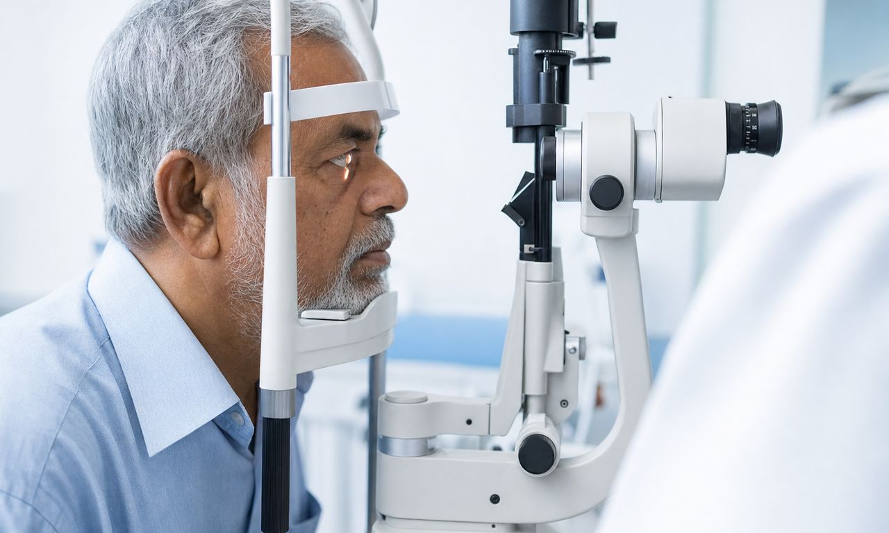
Pseudoexfoliation syndrome (PXF, also called exfoliation syndrome or XFS) is an age-related condition in which a flaky, white protein material builds up inside the eye. It matters for cataract surgery because it weakens the fibers that hold the lens in place, keeps the pupil from dilating well, and raises the risk of pressure problems and glaucoma. A meta-analysis found that eyes with PXF have about twice the odds of posterior capsule rupture or zonular dialysis during cataract surgery (Vazquez-Ferreiro, JCRS 2016). That sounds alarming, but it is the reason surgeons look for PXF before surgery and plan for it. Cataract surgery in a PXF eye is done every day. It is planned differently, not avoided.
If your surgeon just told you that you have pseudoexfoliation, you may never have heard the word before. Most patients have no symptoms from PXF itself. It is found on examination, usually when the pupil is dilated.
What is pseudoexfoliation syndrome?
PXF is a disorder of connective tissue. Cells in the eye produce an abnormal fibrillar material that settles on the front surface of the lens, the edge of the pupil, the drainage angle, and the zonules, the fine fibers that suspend the lens behind the iris. Under the microscope, the deposits look like dandruff on the lens capsule, often in a characteristic ring pattern. Similar material has been found in tissues elsewhere in the body (Naumann, Ophthalmology 1998).
A few features are worth knowing:
- Age. PXF usually appears after age 50, and its prevalence increases markedly with age. It is especially common in Scandinavian populations, but it occurs worldwide (Fontana, Clin Ophthalmol 2017).
- One eye or both. PXF is often asymmetric. In a Minnesota population study, only 25 percent of patients had it in both eyes at diagnosis, and another 29 percent became bilateral within 15 years (Jeng, J Glaucoma 2007).
- Genetics. Variants in the LOXL1 gene are strongly associated with PXF and exfoliation glaucoma (Thorleifsson, Science 2007). However, the high-risk variants are very common: about a quarter of the general population carries two copies. A genetic test cannot tell you whether you will develop PXF, so testing is not used routinely.
- The rest of the body. Some studies link PXF to cardiovascular disease and stroke, but the evidence is mixed and PXF has not been shown to cause heart disease (Chung, Can J Ophthalmol 2018). Your eye surgeon will mention it to your primary care doctor, not treat it as an emergency.
PXF and glaucoma: related, but not the same thing
Pseudoexfoliation syndrome is the deposit disease. Pseudoexfoliative (exfoliation) glaucoma is optic nerve damage that develops in a subset of PXF eyes when the deposits clog the drainage angle and pressure rises. PXF is the most common identifiable cause of glaucoma worldwide (Ritch and Schlötzer-Schrehardt, Surv Ophthalmol 2001).
Having PXF does not mean you have glaucoma, but it raises the odds. In a Finnish cohort, 32 percent of eyes with exfoliation developed glaucoma over 10 years (Puska, J Glaucoma 2002). Among people with high eye pressure, those with PXF were about twice as likely as matched controls to progress to glaucoma (Grødum, Ophthalmology 2005). PXF-related glaucoma also tends to have higher and more variable pressures than ordinary open-angle glaucoma. That is why anyone with PXF needs lifelong pressure and optic nerve checks, before and after cataract surgery.
How PXF changes cataract surgery
Weak zonules
The zonules act like the springs of a trampoline, holding the lens capsule evenly in all directions. PXF deposits weaken them. During surgery, weak zonules can stretch or tear, which can let the lens capsule shift or, in the worst case, let lens material fall back into the vitreous cavity. Signs of weakness can sometimes be seen before surgery: a lens that wobbles slightly when the eye moves (phacodonesis) or a shallower front chamber. Phacodonesis is reported in about 8 to 10 percent of PXF eyes (Fontana 2017).
A pupil that will not open fully
Surgeons work through the pupil, so a wide pupil gives a safer view. In PXF, deposits and degeneration of the iris muscles limit dilation, even with strong drops. A small pupil is also a feature of intraoperative floppy iris syndrome, and the tools surgeons use overlap.
Pressure and inflammation
PXF eyes tend to have more inflammation after surgery and can have pressure spikes, especially when glaucoma is already present. In a series of 1,122 PXF eyes, pressure above 30 mmHg on the first day after surgery occurred in 4 percent of eyes without glaucoma and 17 percent of eyes with PXF glaucoma. The same study found that, on average, pressure was lower after surgery than before, for up to seven years (Shingleton, JCRS 2008).
How surgeons prepare for cataract surgery in a PXF eye
- A careful dilated slit-lamp exam looking for deposits on the lens and pupil edge, the size of the dilated pupil, and any lens wobble. PXF in one eye prompts a close look at the other.
- Gonioscopy and pressure measurement to evaluate the drainage angle, plus optic nerve imaging and visual fields when glaucoma is suspected.
- Optical biometry, for example with the ZEISS IOLMaster 700 or Alcon Argos, and sometimes anterior segment imaging to check lens position and chamber depth.
- A combined plan when glaucoma is present. If you already have glaucoma, your cataract surgeon and glaucoma specialist may recommend combined cataract surgery with MIGS to address pressure at the same operation. That decision belongs to a coordinated cataract and glaucoma evaluation.
Other conditions change the plan too. PXF can coexist with a weak corneal endothelium, and surgeons protect the cornea the same way they do in cataract surgery with Fuchs endothelial dystrophy.
What surgeons do differently in the operating room
Opening the pupil
When drops are not enough, surgeons can add medication inside the eye. Omidria, a combination of phenylephrine and ketorolac added to the irrigation fluid, helps maintain dilation and reduce pain (Omidria prescribing information). Intracameral phenylephrine or epinephrine is another option. Mechanical devices gently stretch and hold the pupil open: the Malyugin ring, a small square ring made by MicroSurgical Technology, or a set of flexible iris hooks.
Supporting the capsule
The central tool for weak zonules is the capsular tension ring (CTR), a thin open ring placed inside the capsular bag. It spreads tension from healthy zonules to weak areas, keeping the bag round and centered. When a section of zonules is missing or very weak, surgeons may use a Cionni modified CTR, which has an eyelet that can be sutured to the wall of the eye, or an Ahmed capsular tension segment, which supports one part of the bag. During the operation, capsule support hooks, such as the Mackool system, can hold the edge of the capsule opening and steady the bag while the cataract is removed.
Gentler technique
Surgeons reduce stress on the zonules with lower fluid flow and pressure settings, careful rotation of the lens, and a dispersive or highly viscous viscoelastic, such as Healon5, to cushion the tissues and hold the space open. Removing the lens cortex gently is part of the same strategy.
The backup plan
Surgeons who operate on PXF eyes plan for a bag that cannot support a standard lens. Options include placing a three-piece lens in front of the capsule (the sulcus), often with the optic captured through the capsule opening, or fixing the lens to the sclera or the iris. Having those options ready, rather than improvising, is much of what planning means. In one series, PXF eyes judged high risk before surgery needed vitrectomy in 15.6 percent of cases, compared with 2.0 percent of other PXF eyes, which shows how well pre-surgery assessment sorts risk (Shingleton, JCRS 2010).
How common are complications?
In published series, PXF clearly raises risk. The 2016 meta-analysis found pooled odds of posterior capsule rupture or zonular dialysis about 2.1 times higher than in eyes without PXF. In the UK Cataract National Dataset, the overall rate of posterior capsule rupture or vitreous loss was 1.92 percent, and pseudoexfoliation or phacodonesis was an independent risk factor (Narendran, Eye 2009).
Two points put those numbers in context. First, most PXF eyes have uncomplicated surgery. Second, many complications arise from surprises, and PXF becomes much less surprising when it is identified before surgery and the devices and backup lens are prepared. Most patients recover on a normal cataract surgery recovery timeline. Infection risk is not meaningfully different, and our guide to endophthalmitis after cataract surgery covers the warning signs every patient should know.
After surgery: what to expect with PXF
Early pressure checks
Because a pressure spike is more likely, especially with glaucoma, surgeons usually check PXF eyes within about a day of surgery and may add pressure-lowering drops. Pain, nausea, or blurred vision that worsens in the first days should prompt a same-day call. Our overview of cataract surgery side effects explains what is normal.
Long-term lens stability
Years after an uncomplicated operation, weakened zonules can occasionally give way and let the lens, still inside its capsule, slip out of position. PXF is the most common condition associated with this. In one series of late in-the-bag dislocations, half of the eyes had PXF, and dislocation occurred on average 8.5 years after surgery (Davis, Ophthalmology 2009). A meta-analysis estimated about six times the odds with PXF (Vazquez-Ferreiro, Acta Ophthalmol 2017). Capsular tension rings probably help but do not fully prevent it. Late dislocation is still uncommon, and it is treatable with surgery to reposition or exchange the lens. Know the symptoms of a dislocated lens after cataract surgery, such as sudden blur or seeing the lens edge.
Ongoing glaucoma monitoring
Cataract surgery does not cure PXF. The deposits continue to form, and glaucoma can develop later even if you did not have it before surgery. Keep regular pressure and optic nerve checks for life.
Can I have a premium IOL if I have PXF?
Sometimes. Toric and multifocal lenses depend on staying centered and on axis, and PXF raises the risk of capsule contraction and decentration over time. A 2017 review advised caution for that reason (Fontana 2017), while a 2024 series reported good toric and multifocal results in selected PXF eyes over about 44 months (Rementería-Capelo, Eur J Ophthalmol 2024). Glaucoma matters too, because multifocal lenses reduce contrast, and our guide to PanOptix, Vivity, and Symfony compared explains how those lenses differ.
You may be a good candidate if:
- Your PXF is mild, the lens is stable on examination, and the pupil dilates reasonably well.
- Your pressure is normal and there is no glaucoma damage.
- Your macula is healthy on OCT and your expectations are realistic.
We would want to talk more if:
- There are signs of moderate zonular weakness, which may favor a monofocal lens with a capsular tension ring.
- You have early, well-controlled glaucoma.
- You have had LASIK or other refractive surgery, or you are very nearsighted. See cataract surgery in highly myopic eyes.
This is probably not the right option if:
- You have advanced exfoliation glaucoma with visual field loss.
- The lens wobbles visibly or has already shifted position.
- A previous operation in the eye involved vitreous loss or capsule damage.
PXF is a reason to choose your surgeon carefully. Experience with pupil expansion devices, capsular tension rings, and backup lens fixation matters more here than in a routine case. Ask your surgeon how often they operate on eyes with weak zonules and what their backup plan is. You can find a fellowship-trained cataract surgeon near you, compare top-rated cataract surgeons by patient reviews and experience, or review common cataract symptoms.
Frequently asked questions
Can I still have cataract surgery if I have pseudoexfoliation syndrome?
Yes. Cataract surgery in PXF eyes is performed routinely. The risk of zonular or capsule problems is higher, roughly double in published series, so surgeons plan ahead with pupil expansion, capsular tension rings, gentle technique, and a backup lens option. Most PXF eyes have uncomplicated surgery and a normal recovery.
What is a capsular tension ring, and will I need one?
A capsular tension ring is a thin, open ring placed inside the lens capsule to spread tension from healthy zonules to weak ones and keep the capsule centered. Many surgeons use one in PXF eyes with signs of zonular weakness. Whether you need one depends on your exam and on what the surgeon finds during the operation.
Does PXF mean I will get glaucoma?
No, but the risk is higher. In a Finnish cohort, 32 percent of eyes with exfoliation developed glaucoma over 10 years. Many people with PXF never develop it. Because pressure can rise at any time, anyone with PXF needs regular pressure and optic nerve checks for life, including after cataract surgery.
Can my IOL fall out of position years after cataract surgery?
It can happen, but it is uncommon. PXF is the condition most often associated with late lens dislocation, which in one series occurred on average about 8.5 years after surgery. It is treatable, usually by repositioning or exchanging the lens. Sudden blur or seeing the lens edge should prompt an exam.
Is PXF hereditary?
Genetics plays a role. Variants in the LOXL1 gene are strongly associated with PXF, and it clusters in some families and populations. However, the risk variants are common in people who never develop PXF, so genetic testing cannot predict it and is not used routinely. Family members should simply have regular eye exams.
Should I see a surgeon who specializes in complex cases?
It is reasonable to ask. Mild PXF is managed by many cataract surgeons. If you have visible lens wobble, a very small pupil, or glaucoma, look for a surgeon experienced with capsular tension rings, pupil expansion devices, and backup lens fixation, and ask how they would handle weak zonules.
This article is general information about cataract surgery, not medical advice, and it is not a substitute for consultation with a qualified ophthalmologist. Read our medical disclaimer.
Choosing a Cataract Surgeon?
Compare 304 top cataract surgeons with real patient reviews, ratings, and locations.
Browse the directory · Search by ZIP · Read the national guide
Latest Articles
Toric IOL for Astigmatism: A Surgeon's 2026 Guide

Same-Day Cataract Surgery on Both Eyes (ISBCS): A Surgeon's 2026 Guide

Cataract Surgery with Glaucoma: How Combined MIGS Procedures Work in 2026